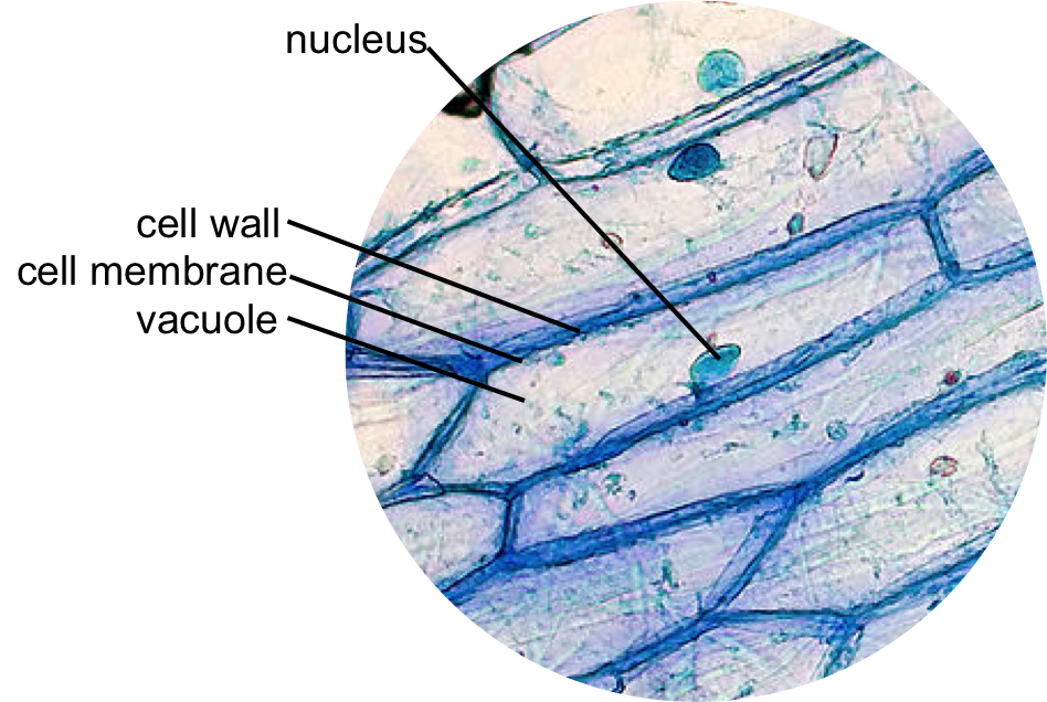
animal cell under microscope labeled
Animal cell below microscope labeled. Here is an electron micrograph of an animal cell with the labels superimposed.

An Electron Micrograph Of A Mouse Liver Cell Dna Learning Center Electrons Cell Learning Centers
When labeled with fluorescent dyes they are invaluable for locating specific molecules in cells by fluorescence microscopy Figure 9-15.

. There are also more intriguing shapes such as curved spherical concave and rectangular. Get more skin-labeled diagrams on social media for anatomy learners. Draw a diagram of one cheek cell and label the parts.
Animal Cell Diagram Under Light Microscope. Our hair grows from follicles located under the skin and has two main parts. This shows a generalized animal cell under a light microscope.
View the leaf under low medium and high power objectives and then draw the cells in Figure 22 along with any organelles you can see. Students know the characteristics that distinguish plant cells from animal cells including chloroplasts and cell walls. Illustrated in Figure 2 are a pair of fibroblast deer skin cells that have been labeled with fluorescent probes and photographed in the microscope to reveal their internal structure.
Click or tap the diagram for a simple labelled version. To make observations and draw scale. The precise antigen specificity of antibodies makes them powerful tools for the cell biologist.
Cell is a tiny structure and functional unit of a living organism containing various parts known as organelles. The shape of animal cells also varies with some being flat others oval or rod-shaped. Students know cells divide to increase their numbers through a process of mitosis which results in two daughter cells with identical sets of chromosomes.
A typical animal cell is 1020 μm in diameter which is about one-fifth the size of the smallest particle visible to the naked eye. However the internal structure and organelles are more or less similar. You should observe the cell membrane nucleus and cytoplasm Observation.
Label the photomicrograph in the activity sheet. Again if you see these chief cells under the electron microscope you will find the abundant endoplasmic reticulum and well-developed Golgi complex. Students know the nucleus is the repository for genetic information in plant and animal cells.
Labeled with electron-dense particles such as colloidal gold spheres they are used for similar purposes in the electron microscope discussed below. Most of the cells are microscopic in size and can only be seen under the microscope. They are all typical elements of a cell.
Hair can be matched by other characteristics that can be viewed under a compound microscope. Within the cell there is a shape of round with a circular structure of granulated part on the epithelial cells. As stated before animal cells are eukaryotic cells with a membrane-bound nucleus.
The cell organelles are enclosed by the plasma membrane including the cell nucleus. Animal cell under the microscope. Add a drop of purple stain specific for animals and cover with a cover slip.
Under the microscope animal cells appear different based on the type of the cell. Add a few drops of a sustainable stain. Differences between animal and human hair.
Skin cells under a microscope. You see that many features are in common. A cell is the smallest functional and structural entity of life that it is easier observing animal cell under light microscope.
Human cheek cells are made of simple squamous epithelial cells which are flat cells with a round visible nucleus that cover the inside lining of the cheekC. You can observe this epithelial animal cell under microscope with high power. As soon as the cells have been mounted and permeabilized on the slide then theyre prepared for staining and viewing.
Observe the cheek cells under both low and high power of your microscope. The animal cell is more fluid or elastic or malleable in structure. Tuesday April 20th 2021.
We all keep in mind that the human physique is amazingly elaborate and one way I discovered to comprehend it is by way of the style of human anatomy diagrams. These are both specific types of cells and from specific species. The granulated area is the cell Cytoplasm while the huge round part is the Nucleus.
So there are numerous possibilities to see the light dark and clear chief cells in the parathyroid gland slide under the light microscope. The plant cell as more rigid and stiff walls. The following labeled drawings must be completed.
Thus these cells sometimes look clear. The nuclei are stained with a red probe while the Golgi apparatus and microfilament actin network are stained green and blue respectively. Heres a diagram of a plant cell.
Generalized Structure of a Plant Cell Diagram. As you can see in the above labeled plant cell diagram under light microscope there are 13 parts namely Cell membrane. Place the leaf on the slide along with a drop of water and then cover with a coverslip.
It also shows the myoepithelial cells that surround each sweat gland of the animal skin. Within the epidermis of a skin you will find squamous diamond-shaped and polyhedral cells under the light microscope. Under a light microscope the cell membrane nucleus and cytoplasm of a cheek cell animal cell can be observed.
Hair under a compound microscope. Be sure to label the chloroplasts the cell membrane and the cell wall.

Cells Microscope Activity Unit Animal Cell Cell Model Cells Project

Cell 8 Pictures Of Plant Cells Under A Microscope Plant Cell Structure Under Microscope Plant And Animal Cells Plant Cell Structure Plant Cell

Histolab4a Htm Histology Slides Science And Nature Animal Cell

Cells And Dna Lesson Science Cells Teaching Biology Teaching Cells

Animal Cell Model Diagram Project Parts Structure Labeled Coloring Plant And Animal Cells Animal Cell Animal Cells Model

Animal Cell Diagram Woo Jr Kids Activities Children S Publishing Cell Diagram Animal Cell Animal Cell Project

Animal Cell Anatomy Banner In 2022 Animal Cell Animal Cell Project Animal Cell Drawing

Draw It Neat How To Draw Animal Cell Animal Cell Drawing Animal Cell Biology Diagrams

Animal Cell Structure And Organelles With Their Functions Jotscroll Animal Cell Organelles Cell Diagram

Google Image Result For Http W3 Hwdsb On Ca Hillpark Departments Science Watts Sbi3u Assigned Work Cell Struct Plant Cell Animal Cells Worksheet Cell Diagram

Printable Labeled And Unlabeled Animal Cell Diagrams With List Of Parts And Definitions Animal Cells Model Animal Cell Cell Diagram

Plant And Animal Cells Revised Animal Cell Project Plant And Animal Cells Plant Cell Project

Muppets Animal Drawing At Paintingvalley Com Explore Collection Of Muppets Animal Drawing Cell Diagram Animal Cell Structure Animal Cell

560 X 364 Pixel Electron Microscope Image Animal Cell And Organelles Labeled Animal Cell Plasma Membrane Organelles

Animal Cell Organelles Sauna Design

Epidermal Onion Cells Under A Microscope Plant Cells Appear Polygonal From The Cell Diagram Plant Cell Diagram Plant Cell

Labeled Animal Cell Diagram Plant And Animal Cells Cell Diagram Animal Cell

Animal And Plant Cells Worksheet Inspirational 1000 Images About Plant Animal Cells On Pinterest Cells Worksheet Animal Cell Plant Cells Worksheet
